Paid Undergraduate Summer Research Program in Biomedical and Veterinary Medical Sciences
Louisiana State University School of Veterinary Medicine (LSU Vet Med) in Baton Rouge, Louisiana, is offering paid 10-week summer research internships to undergraduate and recent post-baccalaureate students. Research topics include toxicology, heart disease, lung biology, immunology, infectious diseases, neuroscience, and more. Students will receive mentorship in research and career planning in a multi-disciplinary and collaborative environment. This opportunity is open to 6 to 10 students annually, depending on funding availability and qualified applicant-mentor matching. Applicants can explore opportunities in any of LSU Vet Med’s three departments: Comparative Biomedical Sciences, Pathobiological Sciences, and Veterinary Clinical Sciences.
Benefits
All participants will receive a $6000 stipend for the 10-week program, university housing, and a meal plan. Some travel expenses may be reimbursed, up to $500. Students will be connected to summer researchers in other colleges/departments within the university through the Office of Undergraduate Research, which creates social programs for summer researchers. All LSU Vet Med summer researchers will receive career mentorship from program mentors and administrators.
Eligibility
All undergraduate students currently enrolled in any U.S. college or university as well as recent postbaccalaureate candidates who are U.S. citizens, permanent residents, or possess student or exchange visas (e.g. F-1, H1, H1B, J1, PR, or TN) are invited to apply. Preference will be given to students with a strong academic background in relevant STEM fields. Applications from students belonging to populations that are traditionally underrepresented in the biomedical field are encouraged. Applications for 2026 program are currently closed.
Application
Interested applicants should visit our website for details. Before you apply, you will need a copy of your unofficial transcript, a personal statement of your research interests, and two letters of recommendation. Identification of potential mentors prior to application is encouraged but not required, but please review our research areas so you can indicate your area of interest. Applications for 2026 program are currently closed.
Research Mentors
Parental administration of third-generation antidepressants and susceptibility to
stress-related disorders Up to twenty percent of pregnant women suffer from depressive
disorders, and approximately three percent are prescribed antidepressants to manage
these conditions. Epidemiological studies have linked prenatal antidepressant exposure
to birth defects, including neonatal adaptive syndrome, while others have reported
cognitive and behavioral disorders associated with untreated maternal depression,
creating a complex paradox that necessitates deeper investigation. Serotonin-norepinephrine
reuptake inhibitors (SNRIs) are third-generation antidepressants that increase monoamine
concentrations in the extracellular space of the brain.
Serotonergic and noradrenergic signaling are critical regulators of stress responses,
and their disruption during development may lead to persistent homeostatic disturbances,
altering behavioral traits and increasing susceptibility to stress-related disorders
such as depression and pathological anxiety. The overarching objective of this study
is to investigate the risks associated with SNRI exposure during early development.
We hypothesize that modulation of neuronal signaling during critical developmental
windows increases the risk of lifelong mental health disorders. To test this hypothesis,
we will: (1) identify stress-related behavioral alterations associated with prenatal
SNRI exposure and determine sensitive developmental windows, (2)
characterize structural and functional changes in stress-related neural circuitry
underlying these behavioral changes, and (3) investigate epigenetic modifications
associated with parental SNRI administration before and after conception. As an undergraduate
researcher, you are expected to contribute to our first objective. The findings from
this study will enhance our understanding of antidepressant-associated neurodevelopmental
risks, inform safer pharmacological interventions for maternal depression, and contribute
to public health policies aimed at mitigating the impact of early-life exposure to
SNRIs.
Summary and hypothesis. Mitochondria regulate immunometabolism and promote host defenses against microbial infections. We recently demonstrated that the formation of mitochondrial-derived vesicles (MDVs) 1, which bud off from mitochondria during infection, enhanced macrophage ability to kill bacteria. However, mechanisms that regulate MDV formation during the immune response remain ill-defined. cardiolipin is a mitochondrial-specific phospholipid that forms membrane curvatures, which contribute to the formation of the inner mitochondrial membrane cristae 2. In addition, cardiolipin acyl chain is modified by the acyltransferase Tafazzin (TAZ) to maintain mitochondrial cristae morphology and functions 3. Importantly, Barth syndrome patients, who have mutations in TAZ, are highly susceptible to bacterial infection 4. Although membrane curvatures are essential for vesicle formation, the role of cardiolipin and acyl chain remodeling for MDV formation and mitochondria-driven host responses has not been investigated. Thus, there is a gap in our understanding of how mitochondria regulate host immune defenses. The overall goal for this project is to determine the importance of cardiolipin and its acyl chain remodeling for the acrophage’s ability to kill bacteria and induce inflammation. Our central hypothesis is that cardiolipin remodeling regulates MDV formation during immune stimulation to promote macrophage antimicrobial functions.
Rationale. Growing evidence supports a role for cardiolipin in the regulation of inflammation during bacterial infection. Importantly, Barth syndrome patients, who have mutations in TAZ, are highly susceptible to bacterial infection 4. Furthermore, in vitro studies suggest that macrophages exposed to the bacterial ligand lipopolysaccharide (LPS) exhibit cardiolipin translocation from inner mitochondrial membrane (IMM) to the outer mitochondrial membrane (OMM) 5. In this model, translocation of cardiolipin to OMM serves as a signaling platform to activate danger immune signaling, like the inflammasomes. Still, the critical role of cardiolipin in mitochondria-derived host defenses, especially during bacterial infection, remains completely unknown.
Approach. To test our hypothesis, we will interfere with cardiolipin biosynthesis and remodeling by knocking down Cardiolipin synthase 1 (Crsl1) and Taz by RNA interference (RNAi) technology, respectively. Comparing these cardiolipin-deficient macrophages with macrophages derived from non-targeted RNAi control, we will investigate the role of cardiolipin in macrophage ability to kill the human bacterial pathogens Salmonella enterica serovar Typhimurium. In addition, we will investigate the role of cardiolipin remodeling in MDV formation, inflammasome activation, and proinflammatory cytokine production during Salmonella infection. We have chosen to focus on Salmonella because Salmonella infection elicits MDV formation, induces strong inflammatory responses, and Salmonella infection is a major global healthcare and economic burden 1,6,7. Our preliminary results suggest that there is a defect in inflammasome activation in Taz knockdown macrophages, which supports a role for cardiolipin remodeling in the regulation of inflammasome activation. Finally, we have developed a method to track membrane-level cardiolipin localization during immune stimulation using a GFP-tagged cardiolipin-binding protein, Stomatin-like Protein 2 (SLP2-GFP), and super-resolution microscopy 8. Using this method, we have observed that cardiolipin-positive MDVs are formed during immune stimulation. Together, these tools will enable us to investigate the contributions of cardiolipin and cardiolipin remodeling to MDV formation, inflammasome activation, and the macrophage ability to kill pathogens. This project will explore cardiolipin localization at a level of resolution that has never been previously achieved. Further, this work will yield mechanistic insight into MDV-dependent antimicrobial defenses. Importantly, our findings will contextualize cardiolipin-dependent innate immune defenses within the framework of a clinically relevant pathogen.
References
- Abuaita, B. H., Schultz, T. L. & O’Riordan, M. X. Mitochondria-Derived Vesicles Deliver
Antimicrobial Reactive Oxygen Species to Control Phagosome-Localized Staphylococcus aureus. Cell Host Microbe 24, 625-636.e5 (2018). - Ikon, N. & Ryan, R. O. Cardiolipin and mitochondrial cristae organization. Biochim. Biophys. Acta Biomembr. 1859, 1156–1163 (2017).
- Acehan, D., Xu, Y., Stokes, D. L. & Schlame, M. Comparison of lymphoblast mitochondria from normal subjects and patients with Barth syndrome using electron microscopic tomography. Lab. Invest. 87, 40–48 (2007).
- Barth, P. G. et al. An X-linked mitochondrial disease affecting cardiac muscle, skeletal muscle and neutrophil leucocytes. J. Neurol. Sci. 62, 327–355 (1983).
- Iyer, S. S. et al. Mitochondrial cardiolipin is required for Nlrp3 inflammasome activation.
Immunity 39, 311–323 (2013). - Nazir, J. et al. Combatting Salmonella: a focus on antimicrobial resistance and the need for effective vaccination. BMC Infect. Dis. 25, 84 (2025).
Substance Use Disorders (SUDs) and Alcohol Use Disorders (AUDs) are major problems in our society today, and there is a great need for better therapies. Repeated substance and alcohol use causes neuroplastic adaptations in the brain through molecular mechanisms. These changes lead to cravings, increased motivation to take the drug, and can cause relapse during times of abstinence. I have studied these lasting effects of abused drugs on the brain since 2009. Almost all of my research experiences have focused on neuroplastic molecular mechanisms that underlie drug-induced behavioral changes in the nucleus accumbens (NAc), a brain area in the mesolimbic dopaminergic pathway vital for reward, stress, and anxiety. I employ cutting-edge viral and chemical tools to manipulate molecular pathways to reverse or mimic drug/alcohol-induced changes in NAc and study their behavioral relevance. The Anderson Lab aims to discover novel, translational treatments for SUDs and AUDs that are capable of reducing drug use by reversing neuroplastic adaptations that occur following chronic use.
Pathogenesis of Pulmonary Diseases: The overall research goal of the Lung Biology Lab is to understand the molecular and cellular mechanisms responsible for neutrophil recruitment, priming, and activation in infected lungs, smoke-exposed lungs, and smoke-exposed lungs and organs followed by infection in the lungs and other organs/tissues. In particular, the Lung Biology Lab is interested in determining the role of pattern recognition receptors (TLRs and NLRs/inflammasomes) and their adaptors with the development of the innate immune response in the lung in murine models. Multiple bacterial pathogens that are studied include: Klebsiella pneumoniae, Streptococcus pneumoniae, Escherichia coli, Pseudomonas aeruginosa, Staphylococcus aureus, and Legionella pneumophila, as causative pathogens of pneumonia. Specific interests of the respiratory disease group include: 1) Delineation of the role of pattern recognition receptors (Toll-like and NOD-like) and neutrophil chemokines, CXCL1, CXCL2, and CXCL5, in lung inflammation and host defense; 2)Elucidation of the mechanisms by which second-hand smoke makes the host susceptible to bacterial infection; 3) Determination of the host defense mechanisms associated with sepsis/septic peritonitis; and 4) Examination of the role of mouse lung-derived stem cells in host protection during bacterial pneumonia. Appropriate gene-deficient mice and human and murine primary cells are currently being used. The ultimate goal of the basic and translation program is to resolve the mechanisms by which these bacterial pathogens cause inflammation in various tissues, and ultimately to design novel treatments and prevention strategies to attenuate inflammation-mediated injury and/or minimize microbial burden.
Pathogenesis of Sepsis: The overall goal of this research is to understand the molecular and cellular immunological mechanisms associated with sepsis. We are particularly interested in innate immune molecules, such as TLRs, NLRs (Inflammasomes) and chemokines in the induction of sepsis.
Seetharama D. Jois
Professor of Cancer Immunology and Computation & Structural Biology
Department of Pathobiological Sciences
School of Veterinary Medicine
Louisiana State University
Skip Bertman Drive
Baton Rouge LA 70803
225-578-9284
sjois@lsu.edu
www.lsu.edu/vetmed/faculty/jois.ph
My research interest is to investigate how proteins interact to control life processes. Proteins interact with one another in highly specific ways, and these interactions play key roles in many cellular processes. In normal cellular processes, these protein interactions are well coordinated to perform cellular functions. Any deregulation of this process can lead to the development of many diseases. My research group is interested in developing peptides/peptidomimetics to modulate protein-protein interactions (PPIs) for therapeutic applications. I use both computational and experimental methods to investigate PPIs and their inhibition. At present, I have three major research projects related to cell-surface proteins and their targeting using peptides, peptidomimetics, and grafted peptides.
Project 1: EGFR-related non-small cell lung cancer (NSCLC) and HER2-positive breast cancer. We design drug-like molecules and evaluate their molecular mechanisms using computational methods, in vitro, and in vivo models.
Project 2: Collaborative research includes developing an imaging agent for EGFR-related colorectal cancer. Imaging agents that emit fluorescence in the near-infrared region are studied using fluorescence microscopy in cells. Furthermore, the photodynamic effect of the molecules is evaluated with lasers at a specific wavelength in 2D and 3D cancer cell culture models.
Project 3: We are also working on multicyclic aptamer peptides with disulfide bonds, resistant to thermal, chemical, and enzymatic degradation, and orally bioavailable. These peptides inhibit co-stimulatory molecules, such as CD2-CD58, which have important implications in autoimmune diseases such as rheumatoid arthritis. Dr. Jois is supported by funding from the National Cancer Institute of the National Institutes of Health.
How does the brain make sense of the world? What happens when it doesn’t? Our lab explores how brain circuits turn sound into perception, thought, and behavior. We use the auditory system as a model to uncover big-picture rules about how neurons communicate across the brain.
Students in our lab get hands-on experience recording electrical signals from neurons and using cutting-edge tools to manipulate these circuits. You’ll see how neural networks actually work! We connect what neurons do with how they’re wired, from individual synapses to entire circuits.
We also study how the brain is affected in conditions such as autism, schizophrenia, and Alzheimer’s disease, linking disrupted brain circuits to real behavioral challenges. Our goal is to understand the brain well enough to help fix what goes wrong.
Why join us this summer? You’ll work with cool tech, ask big questions, build real research skills, and contribute to science that matters. If you’re curious, motivated, and excited about the brain, we’d love to have you.
The Martinez Lab’s research program is focused on the study of vector-borne rickettsial diseases and host-pathogen interactions. The Gram-negative α-proteobacteria of the genus Rickettsia are small (0.3-0.5 x 0.8-1.0 μm), obligate intracellular organisms. These bacteria are transmitted by tick bite inoculation into the skin of the human host and can ultimately damage target endothelial cells especially in the lungs and brain leading to the most severe manifestations of disease, including pulmonary edema and interstitial pneumonia. Although infections are controlled by broad-spectrum antibiotic therapies, untreated or misdiagnosed Mediterranean spotted fever (MSF), Rocky Mountain spotted fever (RMSF) and other spotted fever infections can result in severe morbidity and mortality.
Laboratory efforts are currently aimed at the following research interests: a) characterization of host cell lipid metabolic and catabolic pathways that are utilized by obligate intracellular bacteria to fulfill nutritional requirements during infection of target mammalian cells; b) characterization of essential mammalian cell pathways highjacked by Rickettsia species to establish replicative intracellular niches; and c) determination of genetic determinants in spotted fever group Rickettsia species that contribute to infection and proliferation within endothelial cells, monocytes, macrophages and other mammalian target cells.
Characterization of Guinea Pig Herpesvirus Polyclonal Antiserum: Neutralization Capacity and Temporal Protein Expression Dynamics
Background: Guinea pig herpesvirus (GPHLV) research has been severely limited by the lack of specific reagents. While polyclonal antisera generated against purified virions offer promise for both research applications and therapeutic development, their neutralizing capacity and ability to track viral protein expression kinetics remain uncharacterized. Understanding these properties is essential for advancing GPHLV research and establishing this system as a viable model for herpesvirus pathogenesis.
Methods: We will systematically characterize polyclonal anti-GPHLV antiserum through neutralization assays across multiple dilutions and pre-incubation times. Viral protein expression will be tracked over a 72-hour infection time course using Western blotting to resolve the observed 6-7 band ladder pattern and assign temporal expression classes (immediate early, early, late). Immunofluorescence microscopy will localize viral proteins and assess their subcellular trafficking. Mock-infected controls and pre-immune serum will establish specificity parameters.
Expected Results: We anticipate demonstrating measurable neutralization activity and resolving viral proteins into distinct temporal expression classes. The ladder pattern likely represents different viral glycoproteins and structural proteins with varying molecular weights, providing insights into GPHLV protein processing and maturation.
Impact: This study will validate the first characterized GPHLV-specific reagent, establish standardized protocols for neutralization assays, and provide fundamental data on viral protein expression kinetics. These tools and baseline data will enable future mechanistic studies and antiviral development in this important comparative model system.
Contact
Stephen Costin, Ph.D.
Research and Graduate Education Manager
LSU School of Veterinary Medicine
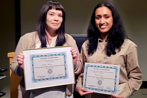
DVM/PhD student Nicole Akers and Anusha Zaman, undergraduate student, received presentation awards at the South Central Chapter of the Society of Toxicology meeting.
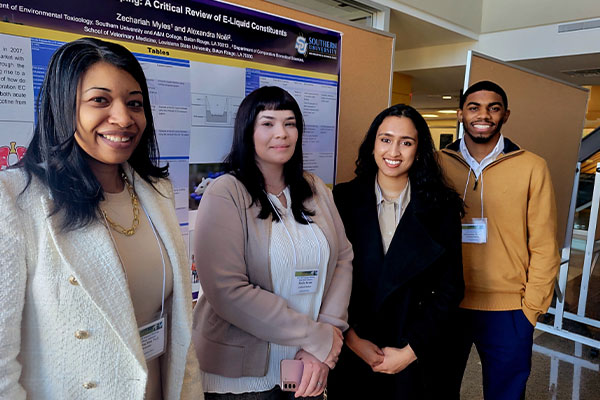
LSU Vet Med researchers attended the South Central Chapter of the Society of Toxicology meeting.